
Common symptoms
- Heavy, tired legs
- Pain or aching in the legs
- Ankle swelling at the end of the day
- Cramps
- Itching
- Bulging veins visible when standing
Centro Médico Teknon · Barcelona
We find the cause of your varicose veins with Doppler ultrasound and choose the minimally invasive technique that suits you best: radiofrequency, endovenous laser, sclerotherapy, microfoam or phlebectomy.
Angiology and Vascular Surgery Unit led by Dr Kerbi Alejandro Guevara Noriega at Centro Médico Teknon.
 Berkelium GroupSurgical Group
Wednesdays and Fridays
09:00–14:00 · 15:00–20:00
Marquesa de Vilallonga 12, floor -1
08017 Barcelona
935 951 710 · Teknon
930 115 874 · Sagrada Familia
What varicose veins are
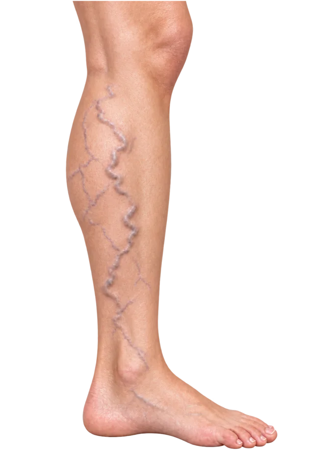
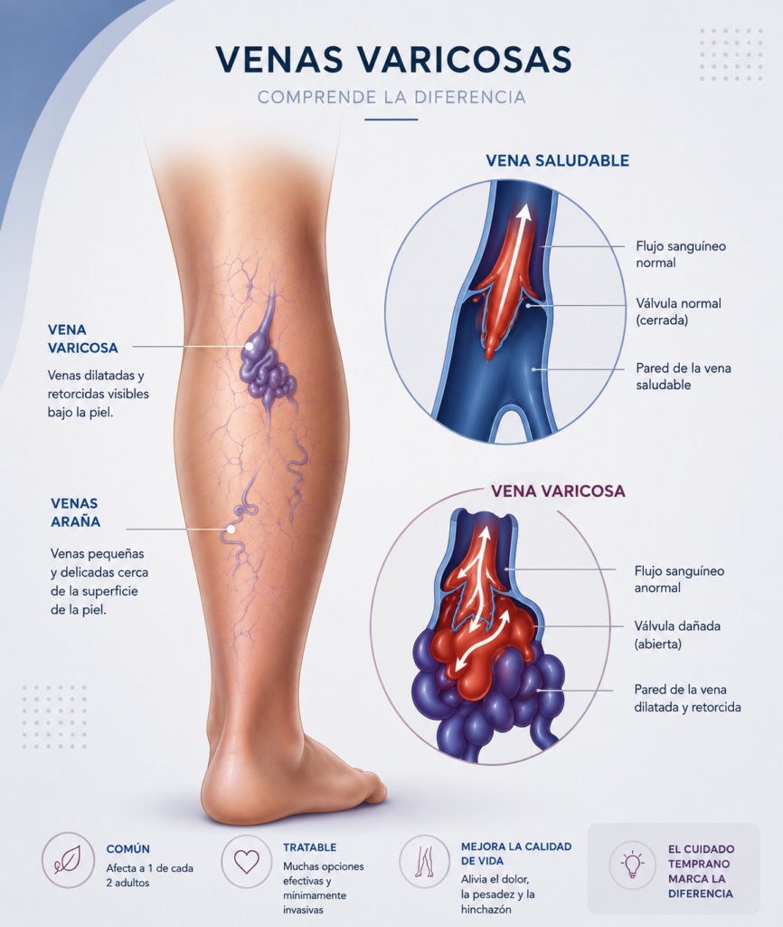
Varicose veins are leg veins that have widened and look twisted under the skin. Many are caused by faulty vein valves: blood that should travel up towards the heart flows backwards, pressure builds and the vein gradually stretches.
That is why not every varicose vein is just a cosmetic issue. Some cause heaviness, pain or swelling and, if venous disease progresses, skin changes or ulcers can develop.
Symptoms
Discomfort tends to get worse at the end of the day or after long hours on your feet.
A vascular assessment is advisable if, as well as varicose veins, you notice:
CEAP classification
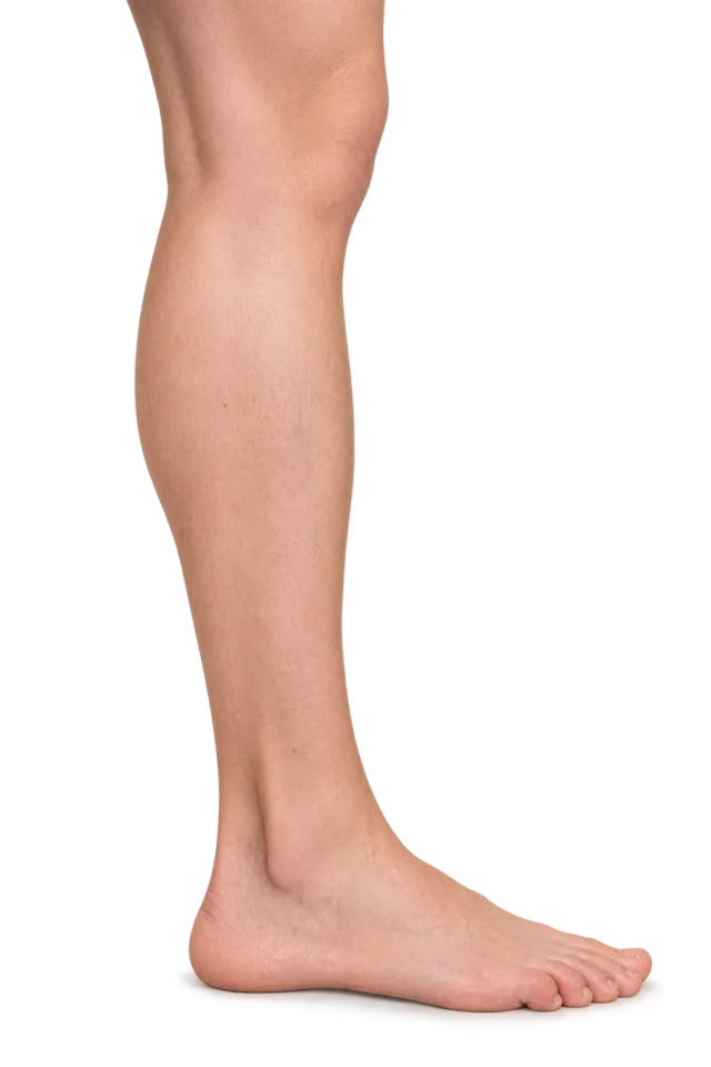
Specialists use the CEAP scale to describe venous disease, from C0 (no signs) to C6 (active ulcer). Varicose veins are stage C2. Slide to see each stage.
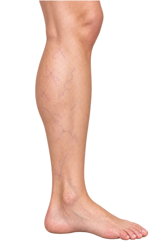
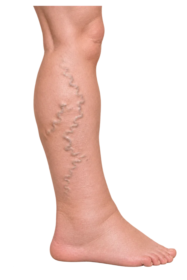
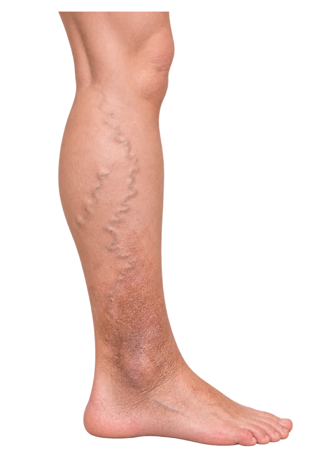
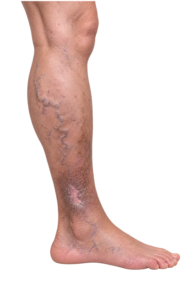
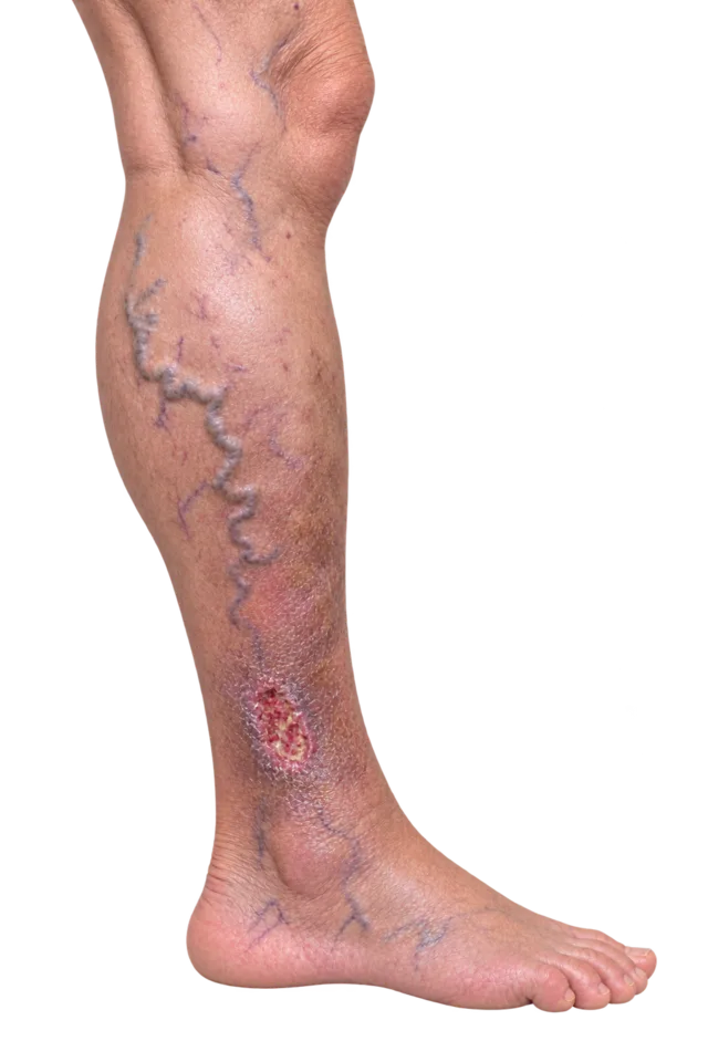
CEAP clinical classification
No visible or palpable signs of venous disease.
CEAP describes the clinical severity of venous disease. It does not represent an inevitable progression.
Diagnosis
Treating varicose veins properly starts with understanding how blood flows in your legs. In clinic we carry out an examination and, when indicated, a venous Doppler ultrasound: a painless scan that shows which veins are failing and whether there is reflux.
This information lets us plan a tailored treatment instead of treating only what is visible on the surface.
Treatments
The technique depends on the size and location of the vein, the degree of insufficiency and your anatomy. Several are often combined.
A catheter delivers controlled heat inside the incompetent saphenous vein to close it.
Learn moreCloses the vein causing the reflux from the inside, without removing it.
Learn moreFine-needle injection for small varicose veins, reticular veins and spider veins. May need several sessions.
Learn moreFoam sclerosant guided by ultrasound for veins that are hard to reach.
Learn moreRemoves surface varicose veins through tiny incisions under local anaesthetic, with minimal or no stitches.
Learn moreFirst the main vein (radiofrequency or laser), then the remaining varicose veins (sclerotherapy or phlebectomy).
Learn moreThere is no one-size-fits-all technique. Sometimes sclerotherapy is enough; in other cases a saphenous vein must be closed first and the remaining varicose veins treated afterwards.
The decision is made with you after the Doppler scan.
Treatment closes or removes the diseased veins, but it cannot stop new ones from appearing over the years: predisposition, age, hormones and the course of venous insufficiency all play a part.
That is why we suggest follow-up visits and preventive measures when they help.
Medical team
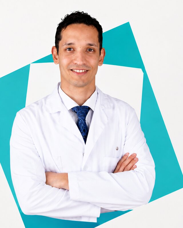
Head of department
Angiology and Vascular Surgery
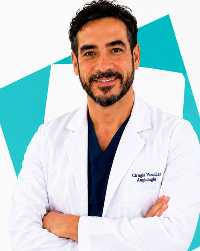
Arterial and aortic disease
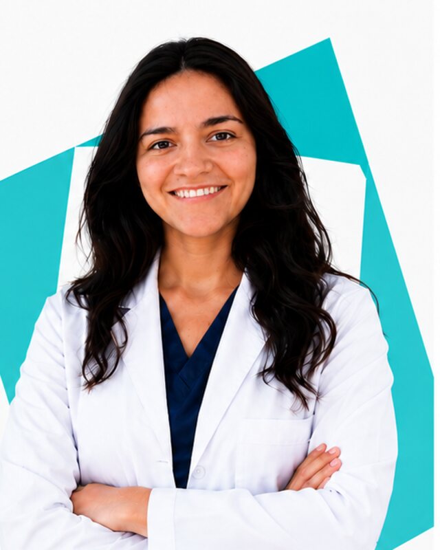
Venous disease
Frequently asked questions
Not always. Some are, but many are caused by venous insufficiency and can lead to heaviness, pain, swelling, skin changes or ulcers. A vascular assessment tells you which.
When indicated, yes. A venous Doppler ultrasound identifies the affected veins, reflux and any blockage, and shows which vein should be treated first.
In many cases, yes, using minimally invasive techniques such as radiofrequency, endovenous laser, sclerotherapy, microfoam or ambulatory phlebectomy under local anaesthetic.
Both close the vein from the inside using a catheter. The choice depends on the venous anatomy, the characteristics of the vein and the team's experience.
It depends on how many veins need treating and how extensive they are. More than one session may be needed.
No. Spider veins are under 1 mm and reticular veins 1–3 mm; veins of 3 mm or more are varicose veins. More about spider and reticular veins.
In consulting room 10, Marquesa Building, Centro Médico Teknon (Marquesa de Vilallonga 12, Barcelona), on Wednesdays and Fridays, 09:00–14:00 and 15:00–20:00. Phone +34 935 951 710.
More on vascular.cat
Sclerotherapy and cosmetic vascular treatment after a prior assessment of the venous system.
Learn moreThrombophlebitis, deep vein thrombosis and post-thrombotic syndrome: individual diagnosis and follow-up.
Learn moreWe identify the cause of the wound and treat the vascular disease that prevents it from healing.
Learn morePelvic congestion, May-Thurner and Nutcracker: embolisation and venous stenting in selected patients.
Learn moreWe explain which technique fits your varicose veins, what recovery involves and what follow-up you will need.